Terms
Aseptic - AntisepticTo work aseptically means that you work in a way that will prevent disease-causing microorganisms to contaminate the material you are working with, without the use of chemical disinfectants. Chemical disinfectants (= germicides) are substances used to inhibit the growth of or to kill microorganisms on either an object or a body surface. An antisepticum is a substance that is used to inhibit the growth of or to kill microorganisms on a body surface. A fungistatic is a substance that kills bacteria. A fungistatic agent (= fungistat) is a substance that inhibits the growth of fungi. A germicide is a substance that kills not only fungi but also other microorganisms. Updated: 2020-12-17. |
|
|
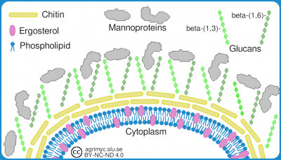
Cellvägg och cellmembranA schematic image of the fungal cell envelope. It is mainly dermatophytes that have mannoproteins. - Click on the image to enlarge it.
IntroductionThe cell wall and the cell membrane are very important structures of fungi because they form the fungal contact surface with the environment. For bacteria, the cell wall and the cell membrane together are usually called the cell envelopes. The same terminology is here used for fungi. Fungi, like other living organisms, have a cell membrane, which consists of a double lipid layer. In fungi, these lipids consist mainly of phospholipids and sphingolipids. The cell membrane of the fungi is thus similar to the cell membrane of mammals, but unlike these, fungi have ergosterol instead of cholesterol in their cell membrane. Many antifungal substances act by inhibiting the synthesis of ergosterol. Like other organisms, the fungi also have proteins in their cell membrane, whichare responsible for transport and biosynthesis etc. However, these are not drawn in the figure. The cell wall consists of a chitin layer, and a layer of glucans (d-glucose polymers). Dermatophytes also have so-called mannoproteins (a type of glycoprotein) on the outside of the cell wall. Updated: 2021-10-14. |
|
|
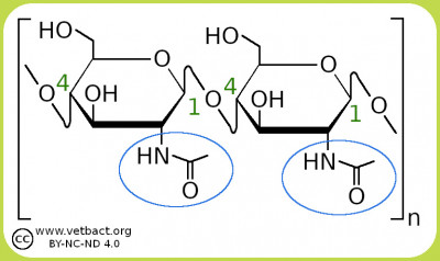
ChitinThe figure shows two N-acetylglucosamine residues, which bind to each other through a β-(1 → 4) linkage. The chitin molecule consists of a long chain of these N-acetylglucosamine residues. The green numbers show the numbering of carbon atoms and the blue circles show the acetyl amine groups. Image: Karl-Erik Johansson (BVF, SLU). - Click on the image to enlarge it. Chitin is a long-chain aminopolysaccharide, which consists of N-acetylglucosamine residues, and is the main component in the cell wall of fungi. Chitin is also included in the shell of arthropods (crustaceans and insects). N-acetylglucosamine residues are also included in the cell wall of bacteria, but not in the form of chitin, but instead in the form of a peptidoglycan, which also contains another polysaccharide (N-acetylmuramic acid) and peptides.The structure of chitin is similar to the structure of another polysaccharide, namely cellulose, which also forms crystalline nanofibrils. Cellulose however, which is the main component of the cell walls of plants, contains hydroxyl groups (-OH) instead of acetyl amine groups. Updated: 2021-05-14. |
|
|
ChytridsChytrides (Chytridiomycota) produce so-called zoospores, which have a flagellum and therefore, they are mobile. Chytrides reproduce mainly asexually through these zoospores, which are produced by mitosis. Chytrides mainly infect algae and other microorganisms, but can also infect higher organisms. Batrachochytrium dendrobatidis, which infects amphibians, and Synchytrium endobioticum, which infects potato plants, are among the chytrides. Updated: 2022-03-24. |
|
|
Club fungiIntroductionClub fungi (Basidiomycota) are filamentous fungi, which include most macroscopic fungi, which in everyday speech are called mushrooms (e.g. chanterelles, boletes trumpet mushrooms, etc.). About 16,000 species have been described in the phylum Basidiomycota. For instance, rust and smut fungi (Class Pucciniomycetes and Class Ustilaginomycetes, respectively), which belong to the club fungi, parasitize cereal crops and cause huge crop losses every year. Sexual reproductionClub fungi have club-like organs (single- or multicellular), called basidia. The word basidium comes from the Greek base, meaning pedestal. Basidia produce sexual meiospores (haploid spores, which are formed by meiosis), which are called basidiospores. Basidia are microscopic and are formed by the terminal cell of the hyphae. Asexual reproductionCertain species of club mushrooms can also multiply asexually by budding. Updated: 2022-03-24. |
|
|
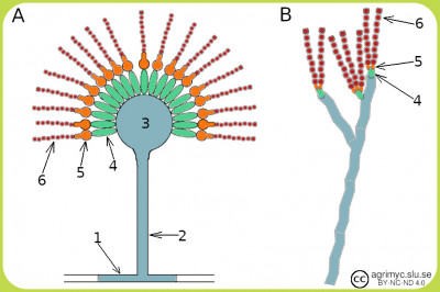
ConidiophoreThe figure shows schematic images of conidiophores of Aspergillus species (A) and Penicillium species (B). Conidiophores consist of the following components: foot cell (1), stalk (2), vesicle (3), metula (4), phialid (5) and conidia (6). Some variation occurs between different species and e.g. A. fumigatus lacks metula. - Click on the image to enlarge it. IntroductionConidiophore is a type of propagating body that produces conidiospores (or conidia) and they are found in e.g. sac fungi (phylum Ascomycota). The conidia are unicellular or multicellular and are formed by lacing from phialid cells (or fialids). This form of reproduction takes place asexually. If the conditions are the right ones, then the conidia form the so-called germination tubes, which grow and form hyphae and fungal mycelium. Updated: 2023-02-08. |
|
|
DermatyphyteFungi, which cause skin, hair and nail infections in humans and animals, are called dermatophytes. Dermatophytes are found among three fungal genera, namely Epidermophyton, Microsporum, Nannizzia (was earlier called Microsporum) and Trichophyton. The dermatophytes get their nourishment by digesting keratin. Keratin is a group of fibrous structural proteins, which occurs in vertebrates and builds up, among other things hair, feathers, nails, claws, hooves and the outer skin layer (epidermis). Dermatophytes usually colonize only the outermost dead layer of the epidermis, because they cannot penetrate the living tissue of immune competent individuals. Dermatophytes have proteases, elastases (serine proteases) and keratinases, which are virulence factors and the degradation products are nutrients for the fungi. These fungi are usually divided into zoophilic, anthropophilic and geophilic dermatophytes according to their normal habitat.
Updated: 2022-11-16. |
|
|
Dimorphic fungiDimorphic fungi can exist in two (or possibly several) different forms and it is usually the temperature that determines in which form the fungus grows. Dimorphic fungi grow in yeast form (small budding form) at 37°C and in mold form (filamentous) at 25°C. There is a memory rule in English for this: Mold in the Cold and Yeast in the Heat. Thus, in mold form, the fungus grows as hyphae. If a dimorphic fungus infects a warm-blooded animal, it will thus grow in yeast form. If the temperature is decisive for the form in which the fungus grows, it is said to be thermally dimorphic. Other factors than the temperature (e.g. the composition of the growth medium) may also affect the form in which the fungus grows. Candida albicans is an example of a dimorphic fungus. Updated: 2021-02-23. |
|
|
Disease typesIntroductionIn both veterinary and human medicine, infectious diseases (caused by fungi, bacteria, parasites or viruses etc.) are usually characterized according to different criteria, and here is one example of classification of diseases within these two disciplines. Classification according to distribution, infectivity and severityEnzootic corresponds to endemic in human medicine and an enzootic disease is a disease, which is always present in a certain animal population, but which at a certain time only affects a small number of animals. Enzootic pneumonia in pigs, caused by Mycoplasma hyopneuminiae, is an example of a bacterial-induced enzootia. Epizootic corresponds to epidemic in human medicine and an epzootic disease, is a serious animal infectious disease that is widespread. Anthrax, which is caused by Bacillus anthracis, is an example of a bacterial epizootic disease. A panzootic is a serious infectious disease, which spreads over large parts of the world and affects one or several species in many countries. Chrytridiomycosis in amphibians is an exaple of a panzootic caused by Batrachochytrium dendrobatidis.The equivalent term in human medicin is pandemic and cholera, caused by the bacterium Vibrio cholerae, gave rise to three pandemics in the 19th century. Zoonosis is a term used in both veterinary and human medicine, as it refers to infectious diseases, which can be transmitted between animals and humans via food, direct contact with infectious animals or through indirect contact with infectious animals via e.g. insect bites. Ringworm is an examples of a zoonosis, which is caused by fungi within the genus Trichophyton. Updated: 2021-03-11. |
|
|
Extremophilic fungiIntroductionExtremophilic fungi are fungi that live in conditions that are considered difficult to survive in for carbon-based life forms. Psychrophiles are organisms that can grow and reproduce at low temperatures (-20°C to +10°C). Thermophiles are organisms that can grow and reproduce at temperatures above +45°C. Among eukaryotic organisms, some fungi are the only thermophiles, which can live and reproduce at temperatures above +45°C. Radioresistant organisms can withstand high levels of ionizing radiation such as UV radiation and nuclear radiation. Xerophiles are organisms that thrive under very dry conditiond (water activity under 0.8). Updated: 2022-12-14. |
|
|
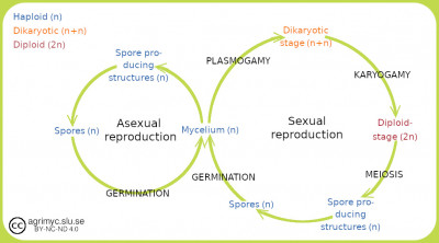
FungiIntroductionThe kingdom of fungi (Fungi) comprises about 150,000 described species, but it is estimated that there are between 1.5 and 3 million species on our planet. The fungi are eukaryotic organisms, i.e. their cells contain a well-defined cell nucleus and in most cases also mitochondria. Fungi are chemoorganoheterotrophic, as are animals. I.e. they use organic carbon compounds both as an energy source and as a carbon source. A unique property of fungi is that the cell wall consists of a mixture of glucans and chitin. Glucans are also found in plants and chitin is the exoskeleton of arthropods, but no other organisms than fungi have a cell wall, which consists of a mixture of these substances. Taxonomy Like bacteria and other living organisms, fungi are hierarchically divided into different taxonomic categories with increasing inclusion ---> genus ---> family ---> order ---> class ---> phylum ---> domain (or kingdom ). For fungi and also certain bacteria there are subcategories, e.g. subkingdom, which then ends up between kingdom and phylum, as well as subphylum, subclass and so on. The classification of fungi has been changed several times as more knowledge becomes available and is likely to be revised further in the near future. The fungi are for the moment divided into 8 different phyla: Ascomycota (sac fungi), Basidiomycota (club fungi), Blastocladiomycota (blastoclads), Chytridiomycota (chytrids), Glomeromycota (arbuscular mycorrhizal fungi), Microsporidiomycota, Neocallimastigomycota and Zygomycota (conjugating fungi). The latter phylum will probably be divided into two different phyla: the Mucoromycota and Zoopagomycota. The phylum Microsporidiomycota may not even belong to the kingdom of Fungi. The figure shows a simplified picture of fungal reproduction by spores, which can be asexual or sexual. Plasmogamy involves fusion of cytoplasm from two cells and karyogami involves fusion of two cell nuclei in the same cell. - Click on the image to enlarge it.
ReproductionFungi multiply by budding or by spores and the spores can be sexual or asexual. The spores thus generally do not have the same function as spores in spore-forming bacteria. Sexual reproduction involves two special stages, plasmogamy and karyogamy. In plasmogamy, the cytoplasm fuses from two different hyphae, which may come from two different fungal individuals (heterotallic fungi) or from the same fungal individual (homotallic fungi). These hyphae will then contain two cell nuclei, which then fuse together through karyogamy and transition to a diploid stage occur. After meiosis, haploid spores can then be produced. DiversityOne reason why there is such great diversity among fungi is that they have managed to adapt to most environments, which occur on our planet. Humans benefit greatly from fungi thanks to:
However, mushrooms are not only good because:
Updated: 2022-05-11. |
|
|
Haploid and diploidIntroductionIn archaea and bacteria, the primary genetic material generally consists of only one circular chromosome. Eukaryotic organisms, on the other hand, generally have several linear chromosomes, which occur in single or double sets. Cells that have a single set of chromosomes (germ cells) are said to be haploid, while cells with a double set (body cells) are said to be diploid. Fungi are eukaryotic organisms and can be haploid, diploid or even tetraploid.
Updated: 2020-12-02. |
|
|
Homothallic and heterothallic fungiIntroductionFungi can multiply both sexually and asexually. Sexual reproduction is common and is the most effective way to achieve genetic variability within a fungal population. Sexual reproduction can take place in two fundamentally different ways, either through self-fertilization or through cross-fertilization. Decisive for this is the type of thallus the fungi have. The term thallus refers to undifferentiated tissue, which in fungi means their mycelium. Self-fertilizationFungi that reproduce by self-fertilization (homothallic fungi) can produce mycelium of both types of reproduction (+ and -) from their own thallus. This can be an advantage because the fungus then does not have to hit another fungus of the opposite type of reproduction. Cross-fertilizationFungi that reproduce by cross-fertilization (heterothallic fungi) can only produce mycelium of one of the reproduction types (+ or -). This means that a heterothallic fungus of a certain species, must encounter another heterothallic fungus of the same species, but of the opposite type of reproduction, in order to be able to reproduce sexually. This is an advantage, as the genetic variability increases and therefore the adaptability of the fungus increases.
Updated: 2021-02-23. |
|
|
Hyphae and pseudohyphaeIntroductionOne usually distinguishes between single-celled and multicellular fungi. Unicellular fungi usually consist of different species of yeast and multicellular fungi of different species of mould. Hyphae are elongated, thread-like and branched filaments, that are made up of tubular cells. Hyphae constitute the mycelium of multicellular fungi, which is the most importande growth mode of these fungi. The growth occurs at the tip of the hypha. Most hyphae have internal cross-walls, which are termed septa (septum sing.) and these hyphae are said to be septated. Some multicellular fungi form hyphae without septa ond these hyphae are termed nonseptated or coenocytic hyphae. Yeasts have pseudohyphae, which consist of cells that have been formed by budding and these cells are connected to the original cell. This collection of cells may be branched. Growth occurs by budding from any cell in the pseudohypha.
Updated: 2020-12-20. |
|
|
ITS sequencesThe figure shows a schematic image of an rRNA cluster in fungi. ITS stands for Internal Transcribed Spacer and IGS for InterGenic spacer. - Click on the image to enlarge it. IntroductionITS sequences means "Internal Transcribed Spacer" sequences and refers to the DNA segments that lie between the rRNA genes in the rRNA operon. Eukaryotic organisms have four different rRNA molecules: 5S, 5.8S, 18S and 26S rRNA or 28S rRNA. The genes for rRNA molecules are organized in clusters or operons and between the genes there are ITS sequences. These clusters are present in many copies in the genome and the figure shows how rRNA genes, ITS sequences and IGS (InterGenic spacer) sequences are organized in the genomes of fungi. Practical useFor the identification and phylogenetic studies of fungi, ITS sequences have proven to be very useful and these sequences are called molecular barcodes. Thanks to the variability in ITS, sequence analysis can provide information about family affiliation and often also species affiliation. Sometimes geographical variants can also be identified, which is useful in epidemiology. In the fungus pages in AgriMyc, links will be provided to ITS sequences deposited in the GenBank database. Fungi have two ITS sequences, while bacteria only have one such gene.
Updated: 2022-05-20. |
|
|
KingdomAccording to an older classification system, the living organisms are divided into five kingdoms: Animalia (animals), Fungi (fungi), Monera (archaea and bacteria), Plantae (plants) and Protista (unicellular organisms with mitochondria, etc.).
This system has been modernized and is now called the Catalog of Life (1), which is abbreviated CoL, and all organisms are there divided into two superkingdoms, Prokaryota and Eukaryota. These two superkingdoms are in turn divided into seven different kingdoms Archaea (archaea or archaebacteria), Bacteria (bacteria or eubacteria), Protozoa (protozoa), Chromista, Fungi (fungi), Plantae (plants), and Animalia (animals). Thus, the first two belong to the superkingdom Prokaryota and the rest belong to the superkingdom Eukaryota. To the kingdom Chromista belong the so-called the water moulds, formerly called fungal-like organisms (or pseudo-fungi). These organisms are now part of the phylum Oomycota, which thus belongs to the kingdom of Chromista. We have chosen to include representatives of phylum Oomycota in AgriMyc because they are important in agriculture and veterinary medicine and because they are often included in fungi chapters in textbooks. ReferenceRuggiero MA, Gordon DP, Orrell TM, Bailly N, Bourgoin T, Brusca RC, Cavalier-Smith T, Guiry MD, 7 & Kirk PM, 2015. A Higher Level Classification of All Living Organisms. PLoS One 10(4): e0119248. Updated: 2021-05-12. |
|
|
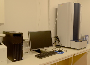
Matrix-Assisted Laser Desorption/Ionization Time Of Flight Mass Spectrometry (MALDI-TOF MS)The instrument in the image belongs to the Department of Biomedical Sciences and Veterinary Public Health at the Swedish University of Agricultural Science (SLU). Lise-Lotte Fernström (+46 18 672389) and Lars Frykberg are responsible for the equipment and it is possible to get samples analyzed. Mass spectrometry based on the MALDI-TOF means that the sample to be analyzed, is adsorbed to some type of carrier material (matrix). The sample is then irradiated with laser UV light, so that the molecules in the sample are broken into charged fragments (ionization), which are thrown towards a detector. The time it takes for the fragment to reach the detector (time of flight) is measured. The time is dependent on fragment size and charge. Also very large molecules (proteins and nucleic acids) can be fragmented and ionized in this way. Large molecules give rise to many fragments and a characteristic mass spectrum, which can be used for identification. More recently MALDI-TOF MS has been used for the identification of microorganisms including yeast and mould. One can perform these analyzes directly on colonies, spores or other suitable material and you will get an analytical response within a minute. The resulting mass spectrum is then compared with stored mass spectra of known relevant microorganisms and the method is considered to be very reliable. The more mass spectra of known microorganisms you have to compare with, the safer the method will be. MALDI-TOF MS is already used in some laboratories for veterinary microbiology and many researchers believe that this technique will be tomorrow's routine method for identification of microorganisms. The instrument is still very expensive, but material costs are low. Reference to a very good review article on applications of MALDI-TOF for identification of fungi: A moldy application of MALDI: MALDI-ToF mass spectrometry for fungal identification. Updated: 2020-12-16. |
|
|
Mycological termsFollowing is a list of various mycological terms in alphabetic order and with brief explanations, which are referred to from the fungus pages. The list will be expanded as new terms appear. If more comprehensive explanations are required, the term gets its own heading in the Termlist. Endophytic means that the organism (fungus or bacteria) can grow into a plant's tissue without causing disease or visible damage. Endophytes have been found in all plants that have been investigated, but the connection between plant and endophyte has not usually been clarified. In some cases, however, a positive effect on the plant has been established. A group of endophytic fungi consists of the so-called the mycorrhizal fungi, which live in symbiosis with e.g. trees. Saprotrophic nutrition means that the fungus, through its hyphae, takes up nutrients (amino acids, fatty acids, disaccharides and glucose), which have been broken down extracellularly (from proteins, lipids, starch or cellulose) by secreting enzymes (proteases, lipases, amylases or cellulases) from the fungus.
Updated: 2023-01-25. |
|
|
MycotoxinsIntroductionMycotoxins are toxic secondary metabolites, which are produced by fungi. They are called secondary metabolites because they are not part of normal growth, development or reproduction. However, secondary metabolites in fungi (eg penicillin) are important for the survival of the fungus in its ecological niche. Mycotoxins can cause disease and death in animals and humans. A fungus can produce several different mycotoxins and a certain mycotoxin can be produced by several different fungal species. The term mycotoxin is usually reserved for substances produced by microscopic fungi, which easily colonize crops, pasture or stored food or feed. Intoxication after intake of contaminated material is termed mycotoxicosis. Mycotxins ar often classified in some major groups. Major groupsAflatoxins are a type of mycotoxins produced by Aspergillus spp. as for instance A. flavus and A. parasiticus. There are four different types of aflatoxins, called B1, B2, G1 and G2. Aflatoxin B1 is most toxic, it is carcinogenic and can cause liver cancer in many different animal species. Aflatoxin B1 is associated with products (e.g. cotton, peanuts, spices, maize and pistachios) from tropical and subtropical regions. Ochratoxins are mycotoxins, which come in three different forms, called ochratoxin A (OTA), OTB and OTC. OTA is the chlorinated form of OTB and OTC is the ethyl ester of OTA. All three forms are produced by Penicillium spp. and Aspergillus spp. A. ochraceus often constitute a contamination of a variety of commodities including wine and beer. A, carbonarius occurs on grapes and when the juice is squeezed out, ochratoxins are released. Ochratoxins are carcinogenic and nephrotoxic. Citrinin occurs in many species of the genus Penicillium and for instance. in P. citrinum. and in some species of the genus Aspergillus. Citrine is nephrotoxic to many species, but it is not known for sure what effect citrine has on humans. Ergot alkaloids are a mixture of toxic alkaloids, which are formed in the sclerotium of some species of the genus Claviceps. These fungi attack various grass species, such as our cereals. If you consume any food that is contaminated by this sclerotium, you can suffer from ergotism, which is a serious disease condition. Patulin is a mycotoxin, produced by species in the genera Aspergillus, Paecilomyces and Penicillium. P. expansum is a species that produces patulin and it often occurs on moldy figs and apples. Patulin is probably not carcinogenic, but it has been shown to damage the immune system in animals. Fusarium toxins are produced by more than 50 species within the genus Fusarium, which infect the grains of growing grains (e.g. wheat and maize). Among the fusarium toxins, fumonisins are especially noticeable, which affect the nervous system in horses and cause cancer in rodents. There are also trichotecenes, which cause chronic and lethal effects in animals and humans, as well as zearalenone, which, however, have not been shown to have any lethal effects on animals or humans. A further number of fusarium toxins have been described (beauvercin, enniatins, butenolide, equisetin and fusarins). Updated: 2021-03-28. |
|
|
Nomenclature of fungiIntroductionNomenclature of fungi refers to naming and fungi and other organisms are named according to the binomial system, which was introduced by Carl Linnaeus (1674-1748). This means that a fungus has both a species name, which is composed of a genus name that tells you to which genus it belongs and a species epithet which, together with the genus name, is unique to the fungus. An example of this is Sporotrix schenkii, where the genus name shows that this fungus belongs to the genus Sporotrix and the species epithet indicates that in this case has the bacterium first been isolated by someone named Schenk. The genus name and the species epithet form together the scientific name of the species, which is always written in italics. Fungal names are international and Latin or latinized Greek are used to form the name. If misunderstandings cannot occur, you can abbreviate the genus name after it has been written for the first time in a text. Macrorhabdus ornithogaster for instance, can be written M. ornithogaster. There are strict international rules for how fungi should be named and these rules have been published in a document named: "International Code of Nomenclature for algae, fungi and plants" (1). In order to get a suggested name for a new fungus accepted, you must register the name in an official register, e.g. MycoBank (2). More information can be obtained through the International Mycological Association (3). Trivial name"Amphibian chytrid fungus" is an example of a trivial name, which is used in English for the fungus Batrachochytrium dendrobatidis, but there are not trivial names for all fungi and sometimes you only use the genus name, which can give rise to misunderstandings. If you are referring to a specific species, it is always best to use the full scientific name. Alternatve species nameFor a long time, there has been a double set of names of fungi, which occur in two different forms (morphs) depending on the sexual stage they are in. Fungi that are in an asexual mitotic stage (morph) are said to be anamorphic and fungi that are in a sexual meiotic stage (morph) are said to be teleomorphic. It was long believed that these stages represented different species and they were consequently given different names. Today, one strives for one and the same name to apply to both morphs and then they talk about holomorph. Subspecies, strains and isolatesSometimes there is a need to divide fungal species into subspecies, because they are too closely related to be considered different species, but too distantly related to be considered as the same species. In this case, a subspecies is introduced by adding a subspecies epithet and writing (subsp. or var.) in front of the subspecies epithet. An example of this is Trichophyton equinum subsp. equinum. When dividing a species into several subspecies, the original species always gets the same subspecies epithet as the species epithet. For fungi, it can be difficult to define a strain because, you may not know if the isolate you have is clonal and then you talk about isolate instead. References1. May TW, Redhead SA, Bensch K et al. 2019. Chapter F of the International Code of Nomenclature for algae, fungi, and plants as approved by the 11th International Mycological Congress, San Juan, Puerto Rico, July 2018. IMA Fungus. doi 10.1186/s43008-019-0019-1 2. MycoBank (Int. Mycol. Assoc.). 3. International Mycological Association (IMA).
Updated: 2022-04-13. |
|
|
Sac fungiIntroductionSack fungi (Ascomycota) or ascomycetes are the largest phylum in the kingdom of fungi (Fungi) and contain about 65,000 described species. These species include i.a. morels, truffles, cap fungi and yeast and mold. The vast majority of fungi, which live in symbiosis with algae and form lichens, also belong to Ascomycota, as well as many veterinarily important fungi. Sexual reproductionWhat characterizes many members of Ascomycota is a microscopic structure for sexual reproduction, called the ascus (meaning sac), where immobile spores (so-called ascospores) are formed. These species are said to be teleomorphic, which refers to the sexual reproduction stage of the fungal life cycle. Asexual reproductionSome species within Ascomycota are asexulla (lack sexual reproduction) and thus do not form ascospores. They are, therefore, called anamorphic species and anamorph is thus an asexual stage of reproduction in the life cycle of the fungi. Asexual reproduction can take place with the help of so-called conidia (= conidiospores), which is a form of spores, which have been formed asexually by mitosis. Mitosis is the form of cell division that gives rise to two genetically identical daughter cells, with the same number of chromosomes as the original cell. By holomorph is meant the entire fungus (all life cycles). The terms anamorph, holomorph and teleomorph are used for sac fungi and basidiomycetes. The concepts have given rise to confusion when it comes to naming fungi, because an anamorphic fungus has been given a certain scientific name, but unfortunately another scientific name is given to the teleomorphic fungus of the same species. On the fungus pages of AgriMyc, one can generally find both names. BuddingAsexual reproduction can also take place through budding and yeasts are unicellular fungi, which often multiply asexually by budding of small "daughter cells". These can sometimes hang on to the mother cell and then form a short chain, which is called a pseudohypha. Updated: 2022-03-17. |
|
|
SclerotiumIntroductionSclerotium can be formed by certain fungal species and consists of a hardened and compact mass of fungal tissue, fungal mycelium and a very small amount of water (5-10%). A sclerotium resembles a cyst with a dark bark-like outer layer. The function is that the fungus should be able to survive under extreme conditions in the form of a sclerotium, which is thus a resting stage, and then begin to grow and thrive again when the conditions return to normal. The fungus can survive during several years in a dry environment in the form of a sclerotium without growing. Examples of fungi, which can form sclerotia
Updated: 2023-01-18. |
|
|
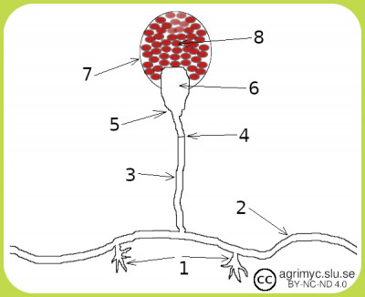
Spores and reproduction of fungiIntroductionSpores in fungi are not the same as spores of bacteria. Fungi use spores (asexual or sexual) to reproduce whereas the bacteria, which form spores, do so to survive adverse conditions (lack of nutrients, extreme pH, high temperature etc). Different fungi form different types of spores and, therefore, different names are used for these spores. However, also fungi can sometimes form spores to survive adverse conditions. Asexual sporesArthrospores (= arthroconidia) are a form of primitive spores, which occur for instance in Coccidiodes immitis. They are formed by fragmentation of hyphae with septa (septated hyphae) and separation of individual hyphal cells. These hyphal cells make up the spores. Blastospores (= blastoconidia) are spores, which are formed by budding from the terminal end of the hyphae. These spores may stay attached to the hyphae for further budding, which will result in a branching chain of blastospores. Ascomyceter and basidiomyceter can produce blastospores. Conidiospores or conidia are formed by so-called conidiophores, which grows aerially from a vegetative hyph. The conidophores branches and at the end of each branch, there is a so-called fialid cell, which produces conidia singly or in chains. Members of the phylum Ascomycota reproduce vegetatively with the help of conidiospores. Chlamydospores can form when conditions are unfavorable for the fungus and then some hyphal cells may lose water and develop a thick cell wall. The cell, which may be at the tip of or in the middle of a hyphae, has then become a chlamydia spore. The chlamydospore can then germinate when the conditions have become favorable again. Ascomycetes and basidiomycetes (for instance Candida albicans and Histoplasma capsulatum) may develop chlamydospores. The figure shows different parts of a sporangium, which grows out of a hyphae (2), which is anchored to the substrate with rhizoids (1). The part closest to the hyphae is called the sporangiophor (3) and it has a septum (4). The part above the septum is called the apophysis (5) and then comes the columella (6) where the sporangiospores (8) are formed. The sporangiospores are surrounded by the wall of thesporangia (7). - Click on image to enlarge it. - Click on the image to enlarge it.
. Sporangiospores are produced in sac-like structures, called sporangia. Sporangia are formed at the end of a hyphae, which grow out of the substrate and are called sporangiophores. The sporangia contain a large number of haploid spores, which are attached to a bladder-like structure called columella. When the sporangia wall ruptures, spores are released into the air. Members of the genera Lichthemia, Mucor, Rhizomucor and Rhizopus produce sporangiospores. Note that the sporangia may have a slightly different structure, depending on which genus the actuall fungus belongs to. Zoospores are unique because they are motile unlike all other types of fungal spores. Zoospores have one single flagellum, which provides mobility, and they are produced in so-called zoosporangia. Members of the phylum Chytridiomycota (chytrides) can produce zoospores. Sexual sporesAscospores are produced by fungi, which belong to the phylum Ascomycota, as the name implies. During sexual reproduction, there must be two different reproduction types (male and female), and one usually talks about plus (+) type and minus (-) type. The hyphal cells and the conidium have N chromosomes each. If a conidium (see above) of the minus-type encounters a plus-type of hyphae, the conidium can "fuse" by plasmogamy with the hyphal cell and form a so-called ascus, which is dikaryotic, i.e. has two cell nuclei (N + N chromosomes). Then the cell nuclei can fuse together through karyogami and then a zygote with a diploid nucleus is formed. The zygote now has two sets of chromosomes (2N) and then genetic material can be exchanged between homologous chromosomes. Then the nucleus in the zygote divides through meiosis and then first the chromosomes (4N) are duplicated and then the nucleus divides twice. This results in 4 new nuclei, each of which has one set of chromosomes. Then the chromosomes are duplicated again and the nuclei now undergo a mitosis, which results in 8 haploid ascospores. Basidiospores are sexual spores, which are produced in a club-shaped organelle, called basidium, by fungi belonging to the phylum Basidiomycota. A typical basidium produces 4 basidiospores exogenously (from the outside) in contrast to ascospores, which are produced endogenously in an ascocarp. Zygospores occur in fungi of the phylum, which was earlier called Zygomycota and which have (or should) be divided into two new phyla (Mucoromycota and Zoopagomycota). Zygospores are diploid and are formed by sexual conjugation between two fungi. Resting sporesTeliospores are thick-walled diploid resting spores that occur in rust and smut fungi and are formed in their fruiting bodies (telia). When the teliospores are mature, they spread to the environment and can rest until conditions become favorable. Then the teliospores germinate resulting in a protomycelium, from which two lines of non-pathogenic, saprophytic and haploid sporidia bud. These sporidia then grow into monokaryotic hyphae, which by conjugation become dicaryotic and can invade the host plant. References1. Fungal spores: Highly variable and stress-resistant vehicles for distribution and spoilage. Updated: 2022-11-16. |
|
|
Temperature dependent fungiIntroductionLike other organisms, different fungi thrive best at different temperatures and the following terms are usually used depending on which temperature range is optimal for growth. Mesophiles are organisms, that can grow and reproduce at temperatures around +37°C (approx. +20 to +45°C). Psychrophiles are organisms that can grow and reproduce at low temperatures (-20°C to +10°C). Thermophiles are organisms that can grow and reproduce at temperatures above +45°C. Among eukaryotic organisms, some fungi are the only thermophiles, which can live and reproduce at temperatures above +45°C. Hyperthermophiles are organisms that can grow and reproduce at temperatures above +80°C. There are no hyperthermophilic fungi. Updated: 2022-12-14. |
|
|
Water moulds (oomycetes)Water moulds (or oomycetes) are a group of eukaryotic microorganisms that resemble microscopic fungi morphologically and by their life style. Therefore, it was previously thought that they belonged to the kingdom of Fungi. Now these organisms have been placed within their own kingdom, namely Chromista, because they differ in several molecular and phylogenetic respects from the actual fungi. For instance the following:
Most water moulds produce two different types of spores. The main spreading spores are asexual and they are motile by their own. They are called zoospores and can move towards or away from a chemical signal, which is called chemotaxis. The chemical signal can consist of for instance substances from potential sources of nutrition. Some water moulds produce asexual spores, which are spread in the air by the wind. Water moulds can also produce sexual spores, which are called oospores. These are translucant, double-walled and spherical structures used to survive in adverse environmental conditions. Water moulds can have a parasitic or saprophytic lifestyle and many of them are important in aquaculture, agriculture and veterinary medicine. Among the algal fungi you will find some of the most notorious plant pathogens and in many text books, these organisms are found under "Fungus-like organisms". The English name water mould (Am. water mold), is inappropriate because most of the oomycetes do not live in water. Updated: 2021-05-20. |
|
|

